

Electron Microscopy
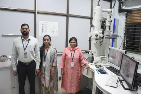
The Electron Microscope Facility at ACTREC is equipped with JEM 1400 PLUS, a Transmission electron microscope (JEOL, Japan) functioning at 120kV with magnification up to 12,00,000 X and 0.2nm resolution. TEM analysis provides a detail ultra- structural information of resin embedded or negatively stained biological samples. The facility extends its services for research and analysis of biological samples, nanoparticles and other applications to the external researchers along with the in-house researchers. The facility is also equipped with instruments such as ultra-microtome, glass knife-maker, auto-trimmer and glow discharge unit.
TRANSMISSION ELECTRON MICROSCOPE Make: JEOL Model: JEM 1400 PLUS
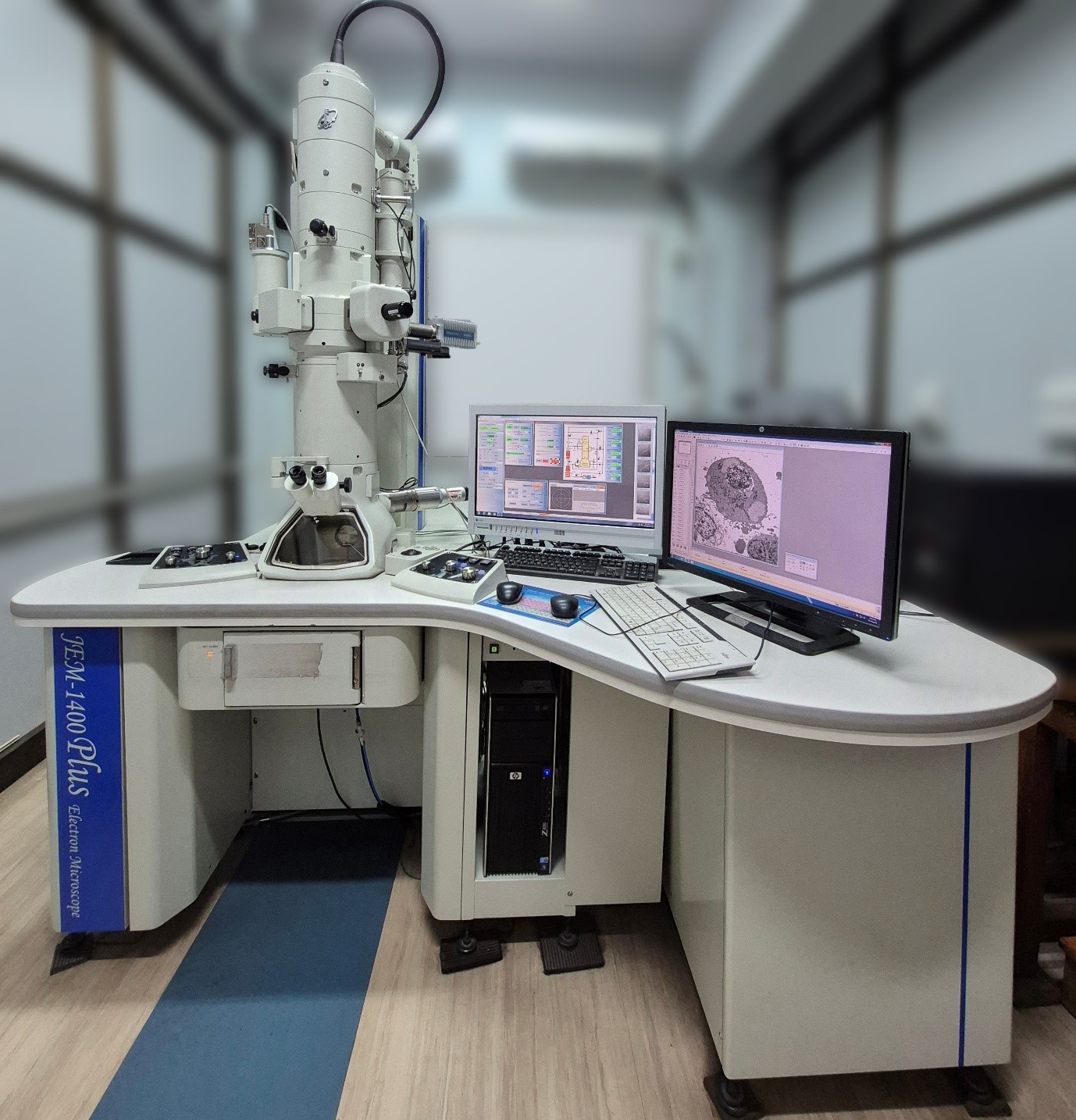
JEM 1400 PLUS is a Transmission electron microscope (JEOL, Japan) functioning at 120kV with magnification up to 12,00,000 X and 0.2nm resolution.
Specifications
| Accelerating voltage | 120 kV |
| Electron Source | Tungsten filament |
| Resolution | HC pole piece |
| Point image | 0.38 nm |
| Lattice image | 0.2 nm |
| Magnification | x200 to x1200000 |
| Camera | Bottom Mounted Olympus TENGRA 5.3 MP CCD Camera |
| Tilt range | ±70° |
Applications
- Imaging of resin embedded cells and tissues.
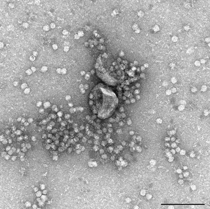
- Imaging of negatively stained small particles (<100nm) like bacteriophages, exosomes, nanoparticles, proteins and DNA.
- Acquisition of immunogold labelled macromolecules to detect specific localization.
- Elemental detection by Bruker EDS system.
EQUIPMENT: ULTRAMICROTOME Make: Leica Model: UC7
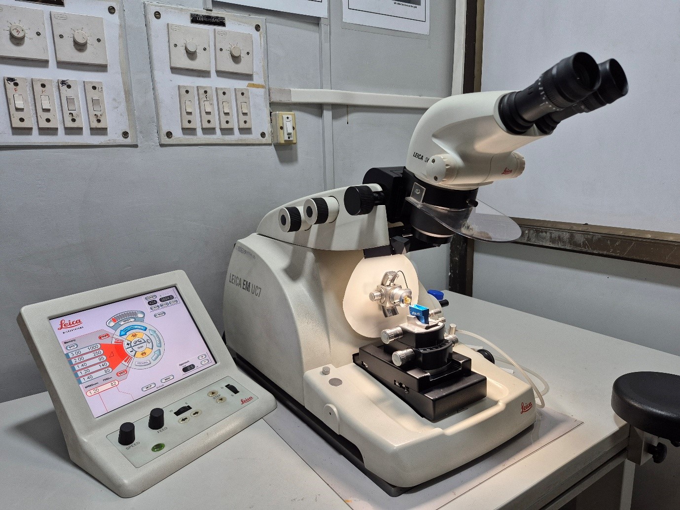
Ultramicrotome provides high-quality preparation of ultra-thin or semi-thin sections for transmission electron microscopy. The desired thickness of the section can be selected with touchscreen control unit and sections can be cut using glass or diamond knives with motorized knife stage. Majorly, 70nm ultra-thin sections are cut for electron microscopy.
Application
- Ultra-thin and semi-thin sectioning of resin embedded cells and tissues.
Additional equipment’s
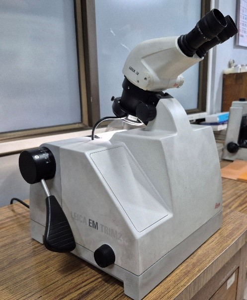 | Leica EM TRIM2 auto-trimmer – for trimming the resin blocks, |
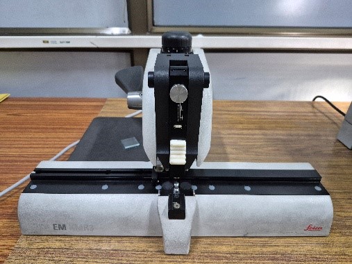 | Leica EM KMR3 – Glass knife maker – for making glass knives. |
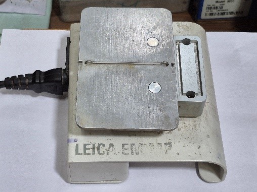 | Leica EM MP Hotplate – for heating the resin sections. |
GLOW DISCHARGE UNIT Make: Ted Pella Model: PELCO easiGlow
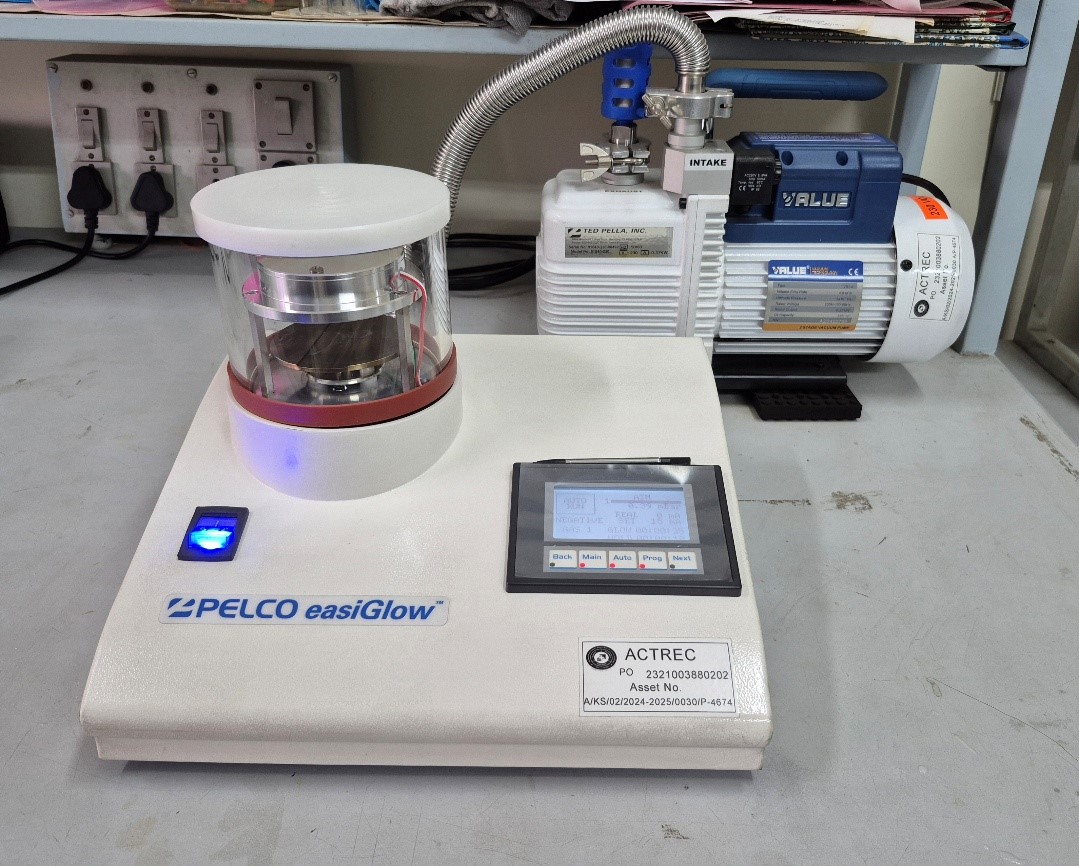
The PELCO easiGlow uses an automated and quick cycle with fully selectable parameters for one of the most common glow discharge applications, making TEM support films or grids hydrophilic using air.
Applications
- To make TEM support films or grids hydrophilic using air
- Cleaning TEM grids
- Adhesion and orientation of proteins, nucleic acids and extra cellular vesicles.
- TEM grid preparation for nanoparticle studies
- The facility carries out sample preparation for routine electron microscopy, including resin block making (solid tissues, monolayer cell cultures, and cell suspensions) followed by ultrathin sectioning using Leica UC-7 ultramicrotome, contrasting, and acquisition.
- The facility also carries out negative staining for small particles (<100nm) like bacteriophages, exosomes, nanoparticles, proteins and DNA.
- The facility provides acquisition of immunogold labelled macromolecules to detect specific localization.
- The facility provides elemental detection by Bruker EDS system
The major areas of electron microscopy analysis are,
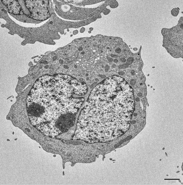
- Ultrastructure study of cell and tissue,
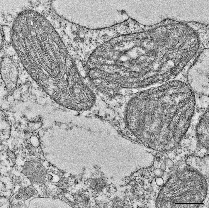
- Studying the details at organelle level like mitochondrial changes, ER - mitochondrial interactions, mitochondrial cristae, cell membrane, nuclear architecture, golgi complexes etc.,
- To check events of autophagy, apoptosis or necroptosis,
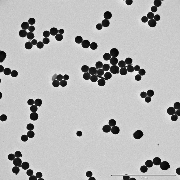
- Characterization of exosomes, bacteriophages and nanoparticles,
- Uptake and effect of nanoparticles / drugs / radiation on cells or tissue,
- EDS analysis of nanoparticles
External users: Please write to the facility regarding your requirement for electron microscopy. The facility will provide the details as per the analysis requirement. The users are requested to discuss over email regarding their study and sample submission followed by booking a slot accordingly. The samples need to be submitted along with a duly filled requisition form. For quotation, a duly filled requisition form can be mailed to the facility email address (tem.facility@actrec.gov.in). For requisition form, please click the link below,
Internal users (ACTREC):
- For routine EM, please check with the facility regarding the sample submission requirements as per the study.
- For negative staining, please book a slot after discussing with the facility for sample submission. Only three samples will be accepted per slot.
Samples should be accompanied by a duly filled requisition form and online requisition.
Requisition form
| ACTREC & DAE | Other than ACTREC institutions | Corporate | ||
|---|---|---|---|---|
| Routine EM | 1000 | 5000 | 10000 | |
| Routine EM + Immunogold labeling | 1500 | 6000 | 12000 | |
| Negative staining | 800 | 4000 | 8000 |
18% GST to be included in addition to the following charges.
Mode of Payment
External users: The users should pay the charges against the quotation or invoice to the following account within a week of the invoice. The users should share the transaction details with the facility once the payment is done. The acquisition will be carried out after the receipt of the payment is confirmed. For e-payment details please click the link below.
Epayment details
Facility-in-charge:
Siddhi A. Redkar
Technical Officer ‘D’
Electron Microscope Facility
(Khanolkar Shodhika - 68)
ACTREC, TMC
Plot No.1 & 2, Sector 22
Kharghar, Navi Mumbai - 410 210.
022-27405000 / 022-68735000
Extension : 5545
Email : tem.facility@actrec.gov.in
EM Staff:
Ms. Arpana Kadam, Scientific Assistant ‘C’
Mr. Shridhar Nadkar, Technician ‘F’
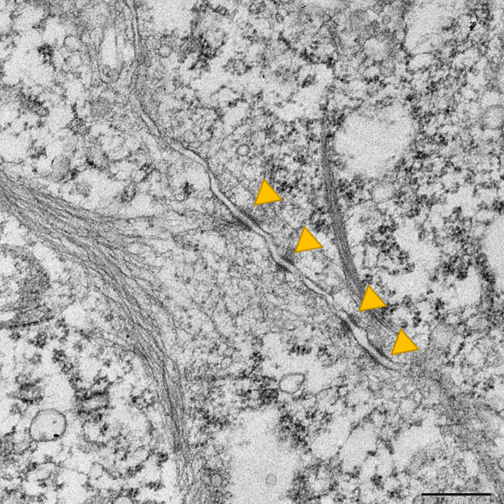 |  |
| Cell to cell Contact | Cell |
 |  |
| Exosomes | Mitochondria |
 | 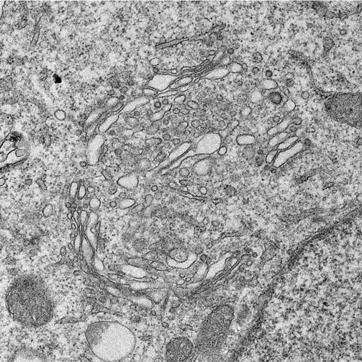 |
| Nanoparticles | Golgi Complex |
2026
- Gupta RJ, Nair H, Sarhadi TR, Nagotu S, Gupta PP, Mishra RM, Dantu SC, Kale A. R5 and R7 positions on fluoroquinolone scaffolds drive F-actin filament disruption. Sci Rep. 2026 Feb 18;16(1):9517.PMID: 41708669; PMCID: PMC13004907.
- Dutta D, Maity A, Gupta SK, Gera P, Jain H, Bagal B, Chatterjee G, Rajpal S, Nayak L, Punatar S, Bandyopadhyay S, Chakraborty A, Chendamarai E, Balasundaram N, Gota V, Khattry N, Sengar M, Mathews V, Hasan SK. Venetoclax induces mitochondrial apoptosis and autophagy to overcome arsenic trioxide resistance in acute promyelocytic leukemia. J Transl Med. 2026 Jan 12;24(1):56.PMID: 41527127; PMCID: PMC12797632.
- Meher RK, Maharana J, Kovvuri J, Kumar KN, Pradhan T, Kailaje V, Ingle A, Sen S, Barkume M, Patkar M, Vaibhaw A, Mir SA, Ghosh A, Kamte S, Kamal A, Kode J. A Phenstatin-based indole-chalcone hybrid as novel anti-inflammatory agent targeting the NLRP3 Inflammasome: Leads from computational and preclinical studies. Bioorg Chem. 2026 Mar;170:109429.Epub 2025 Dec 26. PMID: 41500010.
- Nagare M, Sahoo M, Sengar M, Punatar S, Khattry N, Gokarn A, Bagal B, Jain H, Mirgh S, Epari S, Shet T, Pradhan T, Shirsat S, Barkume M, Mathen C, Gera P, Verma RK, Saldanha E, Chandrani P, Gawde J, Chiplunkar S, Kode J. Defining alteration in bone marrow mesenchymal stem cells (MSC) from acute myeloid leukemia and exploring cultured MSC-conditioned media as a novel anti-leukemia therapy agent. Cancer Immunol Immunother. 2026 Feb 23;75(3):83.PMID: 41729306; PMCID: PMC12929743
- George A, Nair S, Prabhash K, Thakur S, Gera P, Singh A, Chaturvedi P, Rane S, Pradhan T, Sen S, Barkume M, Shetty D, Chaubal K, Ghosh A, Kamte S, Kode J (2026). Generation and characterization of early stage oral cancer cell line of buccal mucosa of Indian origin. Human Cell. 39: 21. PMID: 41410801
- Panigrahi GC, Joshi A, Malhotra D, Gota V (2025). Mitocurcumin induces ROS-/JNK-mediated paraptosis to overcome chemoresistance in non-small cell lung cancer. Naunyn-Schmiedeberg's Archives of Pharmacology. PMID: 41422457.
- Shenoy PS, Dash S, Joshi D, Rekhi B, Gota V, Ray P (2025). Withaferin A exerts cytotoxicity in single/multidrug-resistant gastric and ovarian cancer cells and tumor xenografts through the AKT-NF-κB-STAT3-survivin axis. Molecular Carcinogenesis. 64(12):2037-2051.PMID: 41025926.
- Singh V, Ansari MM, Dutta D, Rajmani RS, Singh A, Singh BN. M. bovis FadR mutant exhibits an altered colony morphotype and increased virulence. Tuberculosis (Edinb). 2025 Dec;155:102689. Epub 2025 Sep 6. PMID: 40939556.
- Chaudhary N, Mandal D, Choudhary B, Patra S, Jain D, Poonia P, Shaikh S, Tekalkar S, Malvankar S, Shivashankar A, Jog E, Pilankar L, Thorat R, Kailje V, Khanna S, Manna S, Khan B, Jadhav A, Sharma K, Ramu S, Sahoo S, Jolly MK, Dalal SN, Sinha S, Nair N, Ruppin E, Lamballe F, Maina F, Verma N (2025). Low-GPX4 drives a sustained drug-tolerant persister state in TNBC by a targetable adaptive FSP1 upregulation. Redox Biology 87: 103864. PMID: 40972157.
- Chaudhary N, Mandal D, Choudhary B, Patra S, Jain D, Poonia P, Shaikh S, Tekalkar S, Malvankar S, Shivashankar A, Jog E, Pilankar L, Thorat R, Kailje V, Khanna S, Manna S, Khan B, Jadhav A, Sharma K, Ramu S, Sahoo S, Jolly MK, Dalal SN, Sinha S, Nair N, Ruppin E, Lamballe F, Maina F, Verma N (2025). Low-GPX4 drives a sustained drug-tolerant persister state in TNBC by a targetable adaptive FSP1 upregulation. Redox Biology 87: 103864. PMID: 40972157.
- Patnaik C, Reddy BPK, Gupta A, Rath S, Neekhra S, Chakrabarty S, Chaudhari P, Asad S, Ingle A, Srivastava R, De A. Non-invasive imaging-guided preclinical assessments reveal high performance photothermal effect of an Au-solid lipid nanomaterial. Nanoscale. 17(46):26658-26677. PMID: 40792496.
- Jaiswal MA, Karn A, Das A, Kumari A, Tiwari S, Dalal SN (2025). 14-3-3ɛ inhibits premature centriole disengagement by inhibiting the activity of Plk1 and Separase. Journal of Cell Science. 138(14):jcs263808. PMID: 40576425.
- Mehta D, Paradkar A, Nayak P, Rekhi B, Mohanty B, Chaudhari P, Waghmare SK. Establishment and molecular characterization of the novel cutaneous squamous cell carcinoma cell line from advanced-stage Indian patient. Human Cell.38 (4):108. PMID: 40413685.
- Joshi P, Bane S, Chaturvedi P, Gera P, Waghmare SK (2025). Establishment and characterization of patient-derived tongue squamous cell carcinoma cell lines. Human Cell. 38(4):102. PMID: 40394426.
- Dahara R, Bhattacharyya D (2025). Intra-Golgi Golgin PpSgm1 and GRIP domain Golgin PpImh1 synergistically mediate Golgi cisternal stacking. Journal of Cell Science.138 (11):jcs263612. PMID: 40270185.
- Anaganti N, Ujaoney AK, Padwal MK, Basu B. Biochemical characterization and functional insights into DNA substrate-specific activities of a unique radiation-inducible DR1143 protein from Deinococcus radiodurans. Int J Biol Macromol. 2025 May;310(Pt 1):143214.Epub 2025 Apr 16. PMID: 40250669.
- Jog E, Jainarayanan AK, Ferlita A, Chakraborty A, Dalwai A, Yahya S, Shivashankar A, Choudhary B, Chandramouli A, Kazi M, Jain D, Khapre N, Akshaya B, Khan B, Gera P, Patil P, Thorat R, Verma N, Sehgal L, Saklani A, Kamat S, Dalal SN, Choudhary N (2025). Inhibiting de novo lipogenesis identifies a therapeutic vulnerability in therapy-resistant colorectal cancer. Redox Biology.79:103458.PMID: 39705849
- Chaudhary N, La Ferlita A, Choudhary BS, Jog E, Kazi M, Yahya S, Dalwai A, Ostwal V, Kumar S, Redkar S, Khapare N, Kailaje V, B A, Gera P, Bal M, Verma N, Thorat R, Saklani A, Sehgal L, Dalal SN (2025). Patient-derived Organoids and Xenografts Uncover Therapeutic Vulnerabilities in Colorectal Signet Ring Cell Carcinomas. Clinical Cancer Research. 31(7):1359-137 PMID: 39879477.
- Arora S, Nagarkar P, D'Souza JS. Recombinant human FOXJ1 protein binds DNA, forms higher-order oligomers, has gel-shifting domains and contains intrinsically disordered regions. Protein Expr Purif. 2025 Mar;227:106622. Epub 2024 Nov 15. PMID: 39549898.
- Shetake NG, Das SK, Kumar A, Pandey BN. Nano-inducer of ferroptosis for targeted chemotherapy of human triple negative breast carcinoma. Biomater Adv. 2024 Jul;161:213868. Epub 2024 Apr 20. PMID: 38677038.
- Meher K, Paithankar H, Hosur RV, Lopus M. Antiproliferative efficacy and mechanism of action of garlic phytochemicals-functionalized gold nanoparticles in triple-negative breast cancer cells. Biomed Mater. 2024 Apr 29;19(3). doi: 10.1088/1748-605X/ad3ff9. PMID: 38682577.
- Radha G, Pragyandipta P, Naik PK, Lopus M. The mode of action of sorafenib in MDA-MB-231 breast carcinoma cells involves components of apoptotic, necroptotic and autophagy-dependent cell death pathways. Exp Cell Res. 2024 Nov 1;443(1):114313. Epub 2024 Oct 30. PMID: 39486634.
- Meher K, Radha G, Lopus M. Induction of autophagy-dependent and caspase- and microtubule-acetylation-independent cell death by phytochemical-stabilized gold nanopolygons in colorectal adenocarcinoma cells. Nanoscale. 2024 Apr 25;16(16):7976-7987. PMID: 38567463.
- Radha G, Pragyandipta P, Naik PK, Lopus M. Biochemical and in silico analysis of the binding mode of erastin with tubulin. J Biomol Struct Dyn. 2025 Aug;43(13):6522-6529. Epub 2024 Feb 20. PMID: 38375607.
- Lonare A, Raychaudhuri K, Shah S, Madhu G, Sachdeva A, Basu S, Thorat R, Gupta S, Dalal SN. 14-3-3σ restricts YY1 to the cytoplasm, promoting therapy resistance, and tumor progression in colorectal cancer. Int J Cancer. 2025 Feb 1;156(3):623-637. doi: 10.1002/ijc.35176. Epub 2024 Sep 6. PMID: 39239852; PMCID: PMC11622004.
- Phadte P, Bishnu A, Dey P, M M, Mehrotra M, Singh P, Chakrabarty S, Majumdar R, Rekhi B, Patra M, De A, Ray P. Autophagy-mediated ID1 turnover dictates chemo-resistant fate in ovarian cancer stem cells. J Exp Clin Cancer Res. 2024 Aug 10;43(1):222.PMID: 39123206; PMCID: PMC11316295.
- Ketkar M, Desai S, Rana P, Thorat R, Epari S, Dutt A, Dutt S. Inhibition of PERK-mediated unfolded protein response acts as a switch for reversal of residual senescence and as senolytic therapy in glioblastoma. Neuro Oncol. 2024 Nov 4;26(11):2027-2043.PMID: 39021199; PMCID: PMC11534322.
- Nagar V, Godambe LP, Newase SK, Tyagi A. Characterization and Genome Analyses of the Novel Phages P2 and vB_AhydM-H1 Targeting Aeromonas hydrophila. Phage (New Rochelle). 2024 Sep 16;5(3):162-172.PMID: 39372357; PMCID: PMC11447392.
- Garg M, Gandhi K, Gera P, Jadhav SM, Mohanty B, Gurjar M, Sandupatla B, Gala R, Chaudhari P, Prasad M, Chinnaswamy G, Gota V. Implications of chronic moderate protein-deficiency malnutrition on doxorubicin pharmacokinetics and cardiotoxicity in early post-weaning stage. Life Sci. 2024 Aug 1;350:122765. Epub 2024 Jun 1. PMID: 38830506.
- Rauniyar S, Pansare K, Sharda A, Singh SR, Saha P, Chilakapati MK, Gupta S. Raman Spectroscopy Revealed Cell Passage-Dependent Distinct Biochemical Alterations in Radiation-Resistant Breast Cancer Cells. ACS Omega. 2023 Feb 3;8(6):5522-5532. PMID: 36816694; PMCID: PMC9933476.
- Dubey S, Mishra N, Goswami N, Siddiqui MQ, Varma AK. Multimodal approach to characterize the tetrameric form of human PML-RBCC domain and ATO-mediated conformational changes. Int J Biol Macromol. 2022 Dec 31;223(Pt A):468-478.Epub 2022 Nov 8. PMID: 36356867.
- Mishra N, Dubey S, Kumari A, Khan MA, Kuligina ES, Preobrazhenskaya EV, Romanko AA, Yadav LR, Sarin R, Imyanitov EN, Varma AK. Structural implications of amyloidogenic rare variants Ser282Leu and Gln356Arg identified in h-BRCA1. Proteins. 2024 Apr;92(4):540-553. Epub 2023 Dec 1. PMID: 38037760.
- Navarange SS, Bane SM, Mehta D, Shah S, Gupta S, Waghmare SK. Epithelial-to-mesenchymal transition status correlated with ultrastructural features, and TP53 mutation in patient-derived oral cancer cell lines. Mol Biol Rep. 2023 Oct;50(10):8469-8481.Epub 2023 Aug 28. PMID: 37639153.
- Ujaoney AK, Anaganti N, Padwal MK, Basu B. Deinococcus lineage and Rad52 family-related protein DR0041 is involved in DNA protection and compaction. Int J Biol Macromol. 2023 Sep 1;248:125885. Epub 2023 Jul 18. PMID: 37473881.
- Chatterjee S, Ganguly A, Bhattacharyya D. Reprogramming nucleolar size by genetic perturbation of the extranuclear Rab GTPases Ypt6 and Ypt32. FEBS Lett. 2024 Feb;598(3):283-301. Epub 2023 Dec 1. PMID: 37994551.
- Dongre HN, Mahadik S, Ahire C, Rane P, Sharma S, Lukmani F, Patil A, Chaukar D, Gupta S, Sawant SS. Diagnostic and prognostic role of protein and ultrastructural alterations at cell-extracellular matrix junctions in neoplastic progression of human oral malignancy. Ultrastruct Pathol. 2022 Sep 3;46(5):476-489. Epub 2022 Sep 1. PMID: 36049041.
- Shukla P, Dange P, Mohanty BS, Gadewal N, Chaudhari P, Sarin R. ARID2 suppression promotes tumor progression and upregulates cytokeratin 8, 18 and β-4 integrin expression in TP53-mutated tobacco-related oral cancer and has prognostic implications. Cancer Gene Ther. 2022 Dec;29(12):1908-1917. Epub 2022 Jul 22. PMID: 35869277.
- Chaudhary N, Joshi N, Doloi R, Shivashankar A, Thorat R, Dalal SN. Plakophilin3 loss leads to an increase in autophagy and radio-resistance. Biochem Biophys Res Commun. 2022 Sep 10;620:1-7. Epub 2022 Jun 22. PMID: 35772211.
- Ud Din Farooqee SB, Christie J, Venkatraman P. PSMD9 ribosomal protein network maintains nucleolar architecture and WT p53 levels. Biochem Biophys Res Commun. 2021 Jul 23;563:105-112. Epub 2021 May 30. PMID: 34077860.
- Sulkshane P, Pawar SN, Waghole R, Pawar SS, Rajput P, Uthale A, Oak S, Kalkar P, Wani H, Patil R, Nair S, Rane P, Teni T. Elevated USP9X drives early-to-late-stage oral tumorigenesis via stabilisation of anti-apoptotic MCL-1 protein and impacts outcome in oral cancers. Br J Cancer. 2021 Aug;125(4):547-560. Epub 2021 Jun 2. PMID: 34079080; PMCID: PMC8367974.
- Rajendra J, Ghorai A, Dutt S. 14-3-3ζ negatively regulates mitochondrial biogenesis in GBM residual cells. Heliyon. 2021 Nov 12;7(11):e08371. PMID: 34825085; PMCID: PMC8605068.
- Bishnu A, Phadte P, Dhadve A, Sakpal A, Rekhi B, Ray P. Molecular imaging of the kinetics of hyperactivated ERK1/2-mediated autophagy during acquirement of chemoresistance. Cell Death Dis. 2021 Feb 8;12(2):161. PMID: 33558461; PMCID: PMC7870816.
- Bose K, Wagh A, Mishra V, Dutta S, Parui AL, Puja R, Mudrale SP, Kulkarni SS, Gai PB, Sarin R. Loss of GSK-3β mediated phosphorylation in HtrA2 contributes to uncontrolled cell death with Parkinsonian phenotype. Int J Biol Macromol. 2021 Jun 1;180:97-111. Epub 2021 Mar 12. PMID: 33716130.
- Khare D, Kumar R, Acharya C. Genomic and functional insights into the adaptation and survival of Chryseobacterium sp. strain PMSZPI in uranium enriched environment. Ecotoxicol Environ Saf. 2020 Mar 15;191:110217. Epub 2020 Jan 27. PMID: 32001422.
- Salunkhe S, Mishra SV, Ghorai A, Hole A, Chandrani P, Dutt A, Chilakapati M, Dutt S. Metabolic rewiring in drug resistant cells exhibit higher OXPHOS and fatty acids as preferred major source to cellular energetics. Biochim Biophys Acta Bioenerg. 2020 Dec 1;1861(12):148300. Epub 2020 Aug 25. PMID: 32858000.
- Kaur E, Nair J, Ghorai A, Mishra SV, Achareker A, Ketkar M, Sarkar D, Salunkhe S, Rajendra J, Gardi N, Desai S, Iyer P, Thorat R, Dutt A, Moiyadi A, Dutt S. Inhibition of SETMAR-H3K36me2-NHEJ repair axis in residual disease cells prevents glioblastoma recurrence. Neuro Oncol. 2020 Dec 18;22(12):1785-1796. PMID: 32458986; PMCID: PMC7746947.
- Sharda A, Rashid M, Shah SG, Sharma AK, Singh SR, Gera P, Chilkapati MK, Gupta S. Elevated HDAC activity and altered histone phospho-acetylation confer acquired radio-resistant phenotype to breast cancer cells. Clin Epigenetics. 2020 Jan 3;12(1):4.PMID: 31900196; PMCID: PMC6942324.
- Roy Chowdhury S, Bhattacharjee C, Casler JC, Jain BK, Glick BS, Bhattacharyya D. ER arrival sites associate with ER exit sites to create bidirectional transport portals. J Cell Biol. 2020 Apr 6;219(4):e201902114. PMID: 32328626; PMCID: PMC7147096.
- Pansare K, Gardi N, Kamat S, Dange P, Previn R, Gera P, Kowtal P, Amin K, Sarin R. Establishment and genomic characterization of gingivobuccal carcinoma cell lines with smokeless tobacco associated genetic alterations and oncogenic PIK3CA mutation. Sci Rep. 2019 Jun 4;9(1):8272. PMID: 31164688; PMCID: PMC6547758.
- Jain BK, Dahara R, Bhattacharyya D. The golgin PpImh1 mediates reversible cisternal stacking in the Golgi of the budding yeast Pichia pastoris. J Cell Sci. 2019 Sep 6;132(17):jcs230672. PMID: 31391238.
- Kirolikar S, Prasannan P, Raghuram GV, Pancholi N, Saha T, Tidke P, Chaudhari P, Shaikh A, Rane B, Pandey R, Wani H, Khare NK, Siddiqui S, D'souza J, Prasad R, Shinde S, Parab S, Nair NK, Pal K, Mittra I. Prevention of radiation-induced bystander effects by agents that inactivate cell-free chromatin released from irradiated dying cells. Cell Death Dis. 2018 Nov 15;9(12):1142. PMID: 30442925; PMCID: PMC6238009.
- Ali M, Sadhu B, Boda A, Tiwari N, Das A, Musharaf Ali SK, Bhattacharya D, Pandey BN, Kumar A. Thorium decorporation efficacy of rationally-selected biocompatible compounds with relevance to human application. J Hazard Mater. 2019 Mar 5;365:952-961. Epub 2018 Nov 13. PMID: 30616306
- Jain BK, Thapa PS, Varma A, Bhattacharyya D. Identification and characterization of GRIP domain Golgin PpImh1 from Pichia pastoris. Yeast. 2018 Aug;35(8):499-506.Epub 2018 May 21. PMID: 29707810.
- Chandwadkar P, Misra HS, Acharya C. Uranium biomineralization induced by a metal tolerant Serratia strain under acid, alkaline and irradiated conditions. Metallomics. 2018 Aug 15;10(8):1078-1088. PMID: 29999065.
- Kolhe N, Zinjarde S, Acharya C. Responses exhibited by various microbial groups relevant to uranium exposure. Biotechnol Adv. 2018 Nov 15;36(7):1828-1846.Epub 2018 Jul 12. PMID: 30017503.
- Sawant S, Dongre H, Ahire C, Sharma S, Jamghare S, Kansara Y, Rane P, Kanojia D, Patil A, Chaukar D, Gupta S, D'Cruz A, Vaidya M, Dongre P. Alterations in desmosomal adhesion at protein and ultrastructure levels during the sequential progressive grades of human oral tumorigenesis. Eur J Oral Sci. 2018 Aug;126(4):251-262. Epub 2018 Jun 15. PMID: 29905981.
- Salunkhe S, Mishra SV, Nair J, Ghosh S, Choudhary N, Kaur E, Shah S, Patkar K, Anand D, Khattry N, Hasan SK, Dutt S. Inhibition of novel GCN5-ATM axis restricts the onset of acquired drug resistance in leukemia. Int J Cancer. 2018 May 15;142(10):2175-2185. Epub 2018 Jan 13. PMID: 29297932.
- Vishal SS, Tilwani S, Dalal SN. Plakoglobin localization to the cell border restores desmosome function in cells lacking 14-3-3γ. Biochem Biophys Res Commun. 2018 Jan 8;495(2):1998-2003. Epub 2017 Dec 15. PMID: 29253567.
- Shanavas A, Rengan AK, Chauhan D, George L, Vats M, Kaur N, Yadav P, Mathur P, Chakraborty S, Tejaswini A, De A, Srivastava R. Glycol chitosan assisted in situ reduction of gold on polymeric template for anti-cancer theranostics. Int J Biol Macromol. 2018 Apr 15;110:392-398. Epub 2017 Nov 22. PMID: 29174361.
- Chauhan DS, De A et al., 2017. NIR light triggered shrinkable thermoresponsive PNVCL nanoshells for cancer theranostics. RSC Advances, 7(70), 44026–34.
- Gurjar M, Raychaudhuri K, Mahadik S, Reddy D, Atak A, Shetty T, Rao K, Karkhanis MS, Gosavi P, Sehgal L, Gupta S, Dalal SN. Plakophilin3 increases desmosome assembly, size and stability by increasing expression of desmocollin2. Biochem Biophys Res Commun. 2018 Jan 1;495(1):768-774. Epub 2017 Nov 14. PMID: 29146182.
- Sawant SS, Dongre H, Ahire C, Sharma S, Kannan S, Mahadik S, Chaukar D, Lukmani F, Patil A, D'Cruz A, Vaidya MM, Dongre P. A nomogram for predicting the risk of neck node metastasis in pathologically node-negative oral cavity carcinoma. Oral Dis. 2017 Nov;23(8):1087-1098. Epub 2017 Jul 5. PMID: 28580710.
- Mukhopadhyay A, Sehgal L, Bose A, Gulvady A, Senapati P, Thorat R, Basu S, Bhatt K, Hosing AS, Balyan R, Borde L, Kundu TK, Dalal SN. 14-3-3γ Prevents Centrosome Amplification and Neoplastic Progression. Sci Rep. 2016 Jun 2;6:26580. PMID: 27253419; PMCID: PMC4890593.
- Sawant S, Dongre H, Singh AK, Joshi S, Costea DE, Mahadik S, Ahire C, Makani V, Dange P, Sharma S, Chaukar D, Vaidya M. Establishment of 3D Co-Culture Models from Different Stages of Human Tongue Tumorigenesis: Utility in Understanding Neoplastic Progression. PLoS One. 2016 Aug 8;11(8):e0160615. PMID: 27501241; PMCID: PMC4976883.
- Sulkshane P, Teni T. BH3 mimetic Obatoclax (GX15-070) mediates mitochondrial stress predominantly via MCL-1 inhibition and induces autophagy-dependent necroptosis in human oral cancer cells. Oncotarget. 2016 Aug 5;8(36):60060-60079.PMID: 28947954; PMCID: PMC5601122.
- Dmello C, Sawant S, Alam H, Gangadaran P, Tiwari R, Dongre H, Rana N, Barve S, Costea DE, Chaukar D, Kane S, Pant H, Vaidya M. Vimentin-mediated regulation of cell motility through modulation of beta4 integrin protein levels in oral tumor derived cells. Int J Biochem Cell Biol. 2016 Jan;70:161-72. Epub 2015 Nov 29. PMID: 26646105.
- Rengan AK, Bukhari AB, Pradhan A, Malhotra R, Banerjee R, Srivastava R, De A. In vivo analysis of biodegradable liposome gold nanoparticles as efficient agents for photothermal therapy of cancer. Nano Lett. 2015 Feb 11;15(2):842-8. Epub 2015 Jan 7. PMID: 25554860.